jbm > Volume 29(1); 2022 > Article
Greenblatt, Shim, Bok, and Kim: The Extracellular Signal-Regulated Kinase Mitogen-Activated Protein Kinase Pathway in Osteoblasts

Abstract

Extracellular signal-regulated kinases (ERKs) are evolutionarily ancient signal transducers of the mitogen-activated protein kinase (MAPK) family that have long been linked to the regulation of osteoblast differentiation and bone formation. Here, we review the physiological functions, biochemistry, upstream activators, and downstream substrates of the ERK pathway. ERK is activated in skeletal progenitors and regulates osteoblast differentiation and skeletal mineralization, with ERK serving as a key regulator of Runt-related transcription factor 2, a critical transcription factor for osteoblast differentiation. However, new evidence highlights context-dependent changes in ERK MAPK pathway wiring and function, indicating a broader set of physiological roles associated with changes in ERK pathway components or substrates. Consistent with this importance, several human skeletal dysplasias are associated with dysregulation of the ERK MAPK pathway, including neurofibromatosis type 1 and Noonan syndrome. The continually broadening array of drugs targeting the ERK pathway for the treatment of cancer and other disorders makes it increasingly important to understand how interference with this pathway impacts bone metabolism, highlighting the importance of mouse studies to model the role of the ERK MAPK pathway in bone formation.

INTRODUCTION

One of the most evolutionarily ancient and well-studied signaling pathways are the group of mitogen-activated protein kinases (MAPKs), which are present in all eukaryotes, including yeast, where MAPKs regulate responses to pheromones, cell integrity, high osmolarity, or other signals.[1,2] While MAPK pathways are ancient, they underwent a major diversification with the development of vertebrate life concurrent with the evolution of skeletal tissues.[3] Accordingly, each of the major MAPK pathways of the conventional group, the extracellular signal-regulated kinase (ERK), p38 and c-JUN N-terminal kinase (JNK) pathways, have been demonstrated to have a key function in promoting bone formation by both osteoblasts [4-8] and also in regulating bone resorption by osteoclasts.[9-13]
In this review, we focus on the contribution of the ERK pathway to osteoblast differentiation. Both ERK isoforms, ERK1 (MAPK3) and ERK2 (MAPK1) are expressed in osteoblast-lineage cells. The distal portion of the ERK signaling cascade displays a stereotypical “wiring” that is largely similar across cell types, with the MAP2Ks MAPK kinase (MEK)1 and MEK2 serving to activate ERK via phosphorylation. As indicated by mouse genetics, though there are differences in biochemical function, ERK1 and ERK2 have partially redundant roles.[14,15] Similar partial redundancy is seen with MEK1 and MEK2, which is most evident in the development of placental defects in either Mek1−/− or Mek1+/−Mek2+/− embryos.[16] Interestingly, a knock-in of the Mek2 protein-coding sequence into the Mek1 locus demonstrated that Mek-associated developmental phenotypes are driven by the total Mek1 and Mek2 gene dosage, with the MEK1 and MEK2 proteins able to substitute for one another. Upstream of MEK1 and MEK2 MAP2Ks, a wide range of kinases, large kinases in the MAP3K family, serve to activate the pathway in a manner that is both stimulus and cell type-specific, with the most classic activator of the ERK MAPK pathway being the RAF family of kinases. However, as discussed below, a wide range of MAP3Ks contribute to ERK activation in osteoblasts.
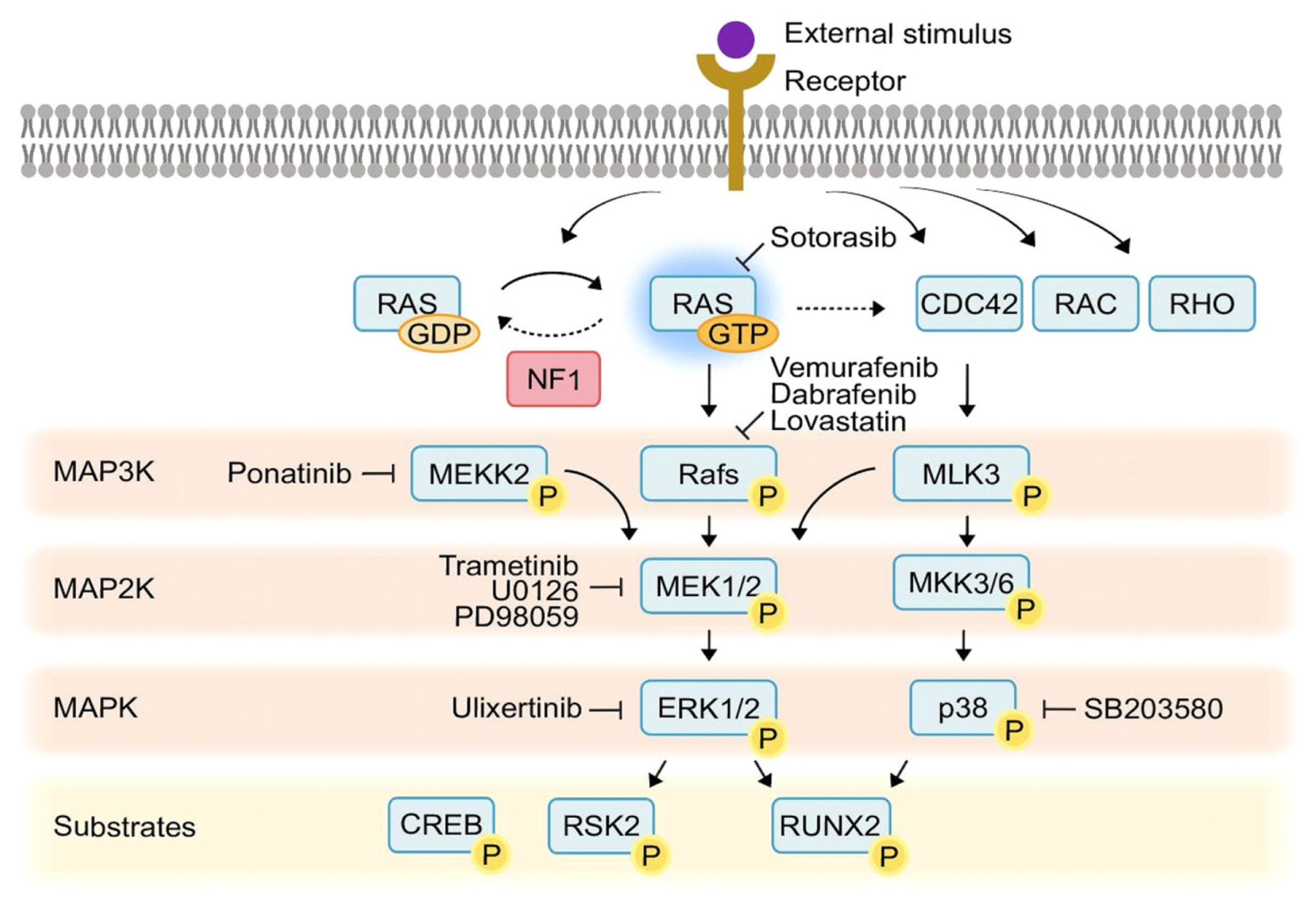
Collectively, mutations in the ERK MAPK pathways are the most common genetic lesions in cancer, and include common recurrent mutations in BRAF and KRAS seen across many tumor types, in addition to other less common mutations in ERK MAPK pathway components such as MEK1 or mutations in upstream signaling components, including EGFR that ultimately lead to ERK activation.[17] Accordingly, this critical importance of ERK in tumorigenesis has led to introduction of a wide range of ERK MAPK pathway inhibitors for treatment of a wide range of tumors, resulting in a powerful pharmacopeia to modulate the ERK MAPK pathway in patients. These drugs include inhibitors of MEK1 and MEK2 such as trametinib,[18] inhibitors of BRAF, a frequently mutated MAP3K upstream of ERK, including vemurafenib and dabrafenib,[19] or inhibitors of mutated KRAS, such as the recently approved sotorasib.[20,21] Due to this, an increasing number of patients are exposed to ERK MAPK pathway inhibitors, and the skeletal consequences of this inhibition must be considered,[22] especially as outcomes and life expectancy for these patients improve and raise the relative importance of long-term sequelae of ERK MAPK pathway inhibition. This development of ERK MAPK pathway inhibitors has also provided a series of potential therapies for skeletal disorders where ERK MAPK pathway overactivation may represent an important pathogenic mechanism, such as neurofibromatosis type 1 (NF1).[23,24] Thus, understanding the role of the ERK MAPK pathway in bone formation by osteoblasts is important both for the intrinsic importance of ERK MAPKs as major regulators of osteoblast differentiation and also due to the wealth of clinically-approved small molecule drugs targeting this pathway (Fig. 1).

PHYSIOLOGIC FUNCTION OF THE ERK MAPK PATHWAY IN OSTEOBLASTS

Most studies on loss-of-function of ERK MAPKs emphasize their critical roles in promoting early commitment and differentiation of skeletal progenitors to the osteoblast lineage and skeletal mineralization (Table 1). For instance, deletion of Erk1/2 in the limb bud mesenchyme and limb skeletal stem cells via Prx1-Cre results in severe hypo-mineralization of the limbs.[25] Similarly, deletion of Mek1/2 in early progenitors committed to the osteoblast lineage using osterix-Cre resulted in impaired bone formation,[8] and mice bearing a transgene of a dominant-negative MEK1 display hypo-mineralization of long bones and the calvarium.[6] While this clearly indicates a role for ERK MAPK in osteoblast differentiation or activity in vivo, somewhat complicating this picture is the observation that ERK MAPK pathway disruption in either Erk1−/−Erk2col2 or Erk1−/−Erk2Prx1 mice leads to defects in the growth plate, specifically an inability to clear and turnover hypertrophic chondrocytes.[25] Given evidence from several investigators that the growth plate cells undergo a trans-differentiation or plasticity process that contributes to the production of marrow-resident osteoblasts,[26-28] this raises the possibility that the blockade in hypertrophic chondrocyte clearance could in turn block the contribution of growth plate cells to the marrow osteoblast pool. Thus, ERK MAPKs could act not only to regulate the differentiation of committed osteoblast lineage cells, but also in pre-commitment progenitors, such as those within the growth plate, to regulate initial commitment to the osteoblast lineage. Notably, growth plate cells appear to only account for a portion of the osteoblasts present in long bones, thus ERK MAPKs may have separate but also important contributions in cathepsin k (CTSK)-lineage periosteal stem cells [29] or leptin receptor (LEPR)+/chemokine (C-X-C motif) ligand 12 (CXCL12)-abundant reticular cells [30-32] or other skeletal stem and progenitor populations.
Interestingly, recent work in a zebrafish model of osteoblast-mediated scale regeneration finds evidence that ERK activation propagates across the entire tissue in a synchronous wave that ultimately coordinates the regeneration process.[33] Evidence from a combination of modeling and proof-of-concept studies using transgenic fibroblast growth factor (FGF) family member expression suggests that this synchronized wave of osteoblast ERK activation is mediated by radial diffusion of an activating ligand, such as FGF, combined with induction of a refractory state in ERK activation after stimulation. This raises the possibility that, if this phenomenon is conserved, ERK MAPKs may not only promote osteoblast activity but mediate broader spatiotemporal coordination of bone formation. Thus, it will be of particular interest to apply a real-time reporter of ERK activity to the bone to examine tissue-wide coordination of skeletal ERK activity in a mammalian system.

LIGANDS ACTING UPSTREAM OF ERK MAPKS IN OSTEOBLASTS

A variety of stimuli activate the ERK MAPK pathway to coordinate signaling during osteoblast differentiation and skeletogenesis. FGF/FGF receptor (FGFR)-mediated signaling regulates osteoblast-lineage commitment, proliferation, maturation, and apoptosis.[34] Numerous in vitro studies demonstrated that the ERK MAPK pathway is required for FGFs-induced osteoblast differentiation through Runt-related transcription factor 2 (Runx2) phosphorylation and activation.[35,36] Genetically, ERK activation is closely linked to the presence of FGFR2 mutations (S252W or P253R) associated with human Apert syndrome.[37-39] Similar ERK activation has also been observed with the craniosynostosis-associated FGFR2 mutation (E731K).[40] A knock-in mouse model carrying a mutation encoding for the P253R alteration in Fgfr2 exhibited ERK activation and premature fusion of the coronal suture, a shortening of the cranial base and long bone growth plates.[37] ERK MAPK pathway inhibition with a MEK1/2 inhibitor partially reversed these phenotypes, including the premature closure of the coronal suture and the growth phenotype in long bones, with these results matching mechanistic studies demonstrating that FGFR2 activation engages the ERK pathway to phosphorylate and stabilize Runx2 via a Peptidyl-prolyl cis-trans isomerase NIMA-interacting 1 (PIN1)-dependent mechanism.[39,41-44]
Insulin-like growth factor 1 (IGF-1) regulates skeletal homeostasis via autocrine and paracrine signaling.[45] Intensive studies showed bone anabolic effects of IGF-1/IGF-1 receptor (IGF-1R) signaling. IGF-1 stimulates mesenchymal stem cell (MSC) proliferation and osteogenic differentiation, and mineralization capacity. Accordingly, Igf-1 overexpression in osteoblasts increased trabecular bone density,[46] whereas IGF-1R deletion in osteoprogenitor cells (Igf1rOsx) or mature osteoblasts (Igf1rOcn) caused delayed mineralization and reduced bone formation.[47,48] ERK activation is required for IGF-1-induced proliferation and osterix expression in human MSCs,[49,50] yet the role of the ERK MAPK pathway in mediating the osteoanabolic effects of IGF-1 signaling in vivo requires further study.
The wingless-type MMTV integration site family member (WNT) signaling pathway also activates ERK MAPK to exert its bone anabolic effect in osteoblast-lineage cells.[47,51] WNT signaling is subdivided into canonical (β-catenin-dependent) and noncanonical (β-catenin-independent) pathways and noncanonical pathway includes MAPK activation, though emerging evidence indicates that the ERK MAPK pathway can also impact canonical signaling. Both in vitro and in in vivo studies showed that WNT3a activates ERK MAPK to induce osteoblast precursor proliferation and differentiation.[52] Likewise, ERK activation suppresses the activity of glycogen synthase kinase 3β, an upstream inhibitor of β-catenin, resulting in bone formation.[51] In addition, mice lacking the WNT receptor Frizzled-9 showed low bone mass without alteration in the canonical WNT/β-catenin signaling pathway, and ERK activation mediated by WNT5a is required for the bone anabolic effect of Frizzled-9 stimulation.[53]
Other upstream stimulators of the ERK MAPK pathway in osteoblasts include the transforming growth factor-β (TGF-β) superfamily, including bone morphogenetic protein (BMP) ligands. These transduce signals to ERK MAPK via the non-SMAD-dependent pathway to regulate stem cell differentiation during bone formation and bone homeostasis.[54] Deletion of TGF-β type I receptor (Alk5) in skeletal progenitors by Dermo1-Cre revealed that TGF-β promotes osteoblast proliferation and early differentiation through a combination of both the SMAD2/3 and ERK MAPK signaling pathways.[55] At the cellular level, TGF-β induces activator protein-1 (AP-1) activation ERK activation.[56] ERK signaling mediates BMP2 or 7-induced osteogenesis and Runx2 expression, whereas constitutive activation of ERK signaling decreased BMP2-induced Runx2 expression and activation.[57,58] BMP2 stimulation leads to Runx2 acetylation and stabilization, potentiating Runx2 transcriptional activity.[59] However, further study is needed to clarify the degree to which the ERK pathway specifically accounts for the activity of BMP stimulation during skeletogenesis.

MAP3KS ACTIVATING THE ERK MAPK PATHWAY IN OSTEOBLASTS

While RAF isoforms (ARAF, BRAF, and c-RAF/RAF1) are classical MAP3Ks that serve to activate MEK1 and MEK2 MAP2Ks, particularly in a variety of oncologic contexts, it is clear that a broader diversity of MAP3Ks function upstream of ERK MAPKs in osteoblasts, often in combination with concurrently activating other MAPK pathways. In particular, studies of BRAF using a Col2-Cre line that targets a wide range of skeletal cells find that the knockout mice have grossly normal initial skeletogenesis.[60] Similarly, deletion of Braf with the osteoblast-targeting Col1-Cre did not produce histologically evident changes in bone architecture in 2-week old mice. Mice with a conditional deletion of c-Raf/Raf1 using Col2-Cre have also been examined, finding impaired clearance of hypertrophic chondrocytes in the growth plate, a finding that may be expected to alter the production of growth-plate derived osteoblasts, though this has yet to be examined.[61] It will be of particular interest to expand and update these findings to see whether RAF isoforms have a more subtle role in regulation of adult bone homeostasis and whether redundancy might be masking the contribution of RAF to osteoblast function.
Given this negative initial data with RAF isoforms, there has been interest in identifying other MAP3Ks that serve to activate ERK MAPKs in osteoblasts. MLK3 (MAP3K11) serves to activate both p38 and ERK downstream of BMP stimulation, and activation of MLK3 induces Runx2 phosphorylation at a combination of p38- and ERK-mediated sites.[62] MLK3 acts downstream of the guanosine triphosphatase (GTPase) CDC42, binding through a CDC42/Rac interactive binding (CRIB) region in MLK3. Accordingly, mutation of the CRIB region phenocopies the skeletal abnormalities of an MLK3 null allele. CDC42 is itself activated by the GEF faciogenital dysplasia 1 (FGD1).[63] As FGD1 is mutated in the human skeletal dysplasia disorder FGD, defective MLK3 signaling is likely a key pathogenic mechanism for this disorder. This also provides a clear example of how impaired ERK signaling in osteoblasts can serve as a basis for human skeletal diseases.
MEKK2 (MAP3K2) is a member of the MAP3K family, and early in vitro biochemical studies have shown that MEKK2 has the capacity to activate downstream MAPK pathways including ERK1/2, JNK, p38, and ERK5.[64-67] Studies of MEKK2 in osteoblasts demonstrated that MEKK2 promotes osteoblast activity by both mediating a nonclassical pathway for deubiquitination and stabilization of β-catenin.[68] MEKK2 has also been suggested to regulate the activation of the JNK pathway downstream of SMURF1 loss-of-function in osteoblasts.[69] However, later studies in MEKK2 deficient osteoblasts failed to find a clear contribution to JNK activation, at least under basal conditions, so the contribution of SMURF1 regulation of MEKK2 activity to osteoblast differentiation and activity remains unclear.[4,68] In keeping with these important functions of MEKK2, MEKK2-deficient mice display severe cortical and trabecular osteopenia alongside calvarial hypomineralization.[68] Recent findings additionally demonstrate that MEKK2 is a key mediator of the aberrant ERK activation occurring in osteoblasts downstream of loss of function of NF1, a GTPase-activating protein that negatively regulates RAS activation.[70] Despite MEKK2-deficient mice displaying severe osteopenia, either genetic or pharmacologic inhibition of MEKK2 prevented the constitutive ERK activation occurring in models of skeletal NF1 disease and overall ameliorated the NF1-associated skeletal phenotypes. This work, alongside several prior studies establishing the ERK MAPK pathway as one, though likely not the only, mediator of NF1-associated skeletal diseases,[23,24,71] argues that ERK MAPKs are not unconditionally pro-anabolic. Rather, there is likely a window of activity that is pro-anabolic, with degrees of activation outside of this range either not promoting osteoblast differentiation or having a deleterious effect. This “window effect” may also have an etiology in the distinct sets of downstream substrates for the ERK MAPK pathway that appear in early versus late-stage osteoblasts, with overactivation of ERK perhaps prolonging ERK activity during the differentiation process and engaging distinct sets of effectors in mature osteoblasts. Ultimately, further study on the context-dependent roles of the ERK MAPK pathway will be needed to determine the basis for this apparent window in ERK activity that allows for bone anabolism. Additionally, a comparison of these studies on MEKK2 and MLK3 emphasizes how each function in a distinct biochemical context, functioning downstream of different upstream activators in a selective manner. This reinforces the general concept that MAP3Ks serve as key integration points that activate downstream MAPK pathways in a manner that is context and cell-type specific. This specificity also suggests that MAP3Ks are a promising point to modulate ERK MAPK signaling while also minimizing the toxicity associated with broad alterations of this essential pathway across multiple tissues.

REGULATORS OF THE ERK MAPK PATHWAY IN OSTEOBLASTS

The NF1 protein negatively regulates RAS MAPK pathway through its GTPase activity. Loss of function NF1 mutations in humans causes NF1, a prevalent genetic disorder with an incidence of approximately 1/3500.[72] NF1 syndrome is characterized by cutaneous neurofibromas and neural tumors, but also a wide variety of non-tumor manifestations including skeletal dysplasia.[73,74] Patients with NF1 display skeletal abnormalities including craniofacial dysmorphogenesis, osteopenia/osteoporosis, short stature, and impaired fracture healing.[75,76] Similarly, conditional mouse models lacking NF1 in various skeletal lineage cells display characteristic features of the skeletal abnormalities of NF1 patients, including impaired bone structure and severe cortical bone porosity in Nf1Prx1,[77] Nf1Col,[71] Nf1Osx,[78] and Nf1Dmp1,[70] mice. To understand the pathogenesis of NF1 skeletal manifestations, several studies have investigated target pathways mediating the aberrant ERK effects of NF1 loss of function in skeletal lineage cells. It has been reported that RAS inhibition by lovastatin attenuates the constitutive activation of ERK MAPKs in osteoblasts isolated from Nf1Col2 mice.[71] NF1-deficiency in bone-forming cells causes a RAS/ERK-dependent increase in the endogenous inhibitor of mineralization, inorganic pyrophosphate, ultimately leading to a bone mineralization deficit.[78]
Interestingly, altered ERK MAPK pathway regulation can result in divergent skeletal phenotypes. Protein-tyrosine phosphatase, nonreceptor-type 11 (PTPN11), also known as SHP2, is a SH2 domain-containing non-receptor protein tyrosine phosphatase that acts to regulate ERK activity in cell type and context-dependent manner, particularly downstream of receptor tyrosine kinase stimulation.[79] As with NF1, while ERK is a prominent downstream mediator of PTPN11 it is not the only downstream target, and other pathways, such as nuclear factor of activated T cells calcium signaling or PKA signaling can also be regulated.[80,81] Gain-of-function mutations in PTPN11 enhance ERK signaling in osteoblasts and are seen in approximately 50% of patients with Noonan syndrome, which is associated with skeletal manifestations including impaired skeletal mineralization with onset in childhood and short stature.[82-84] Interestingly, loss of function in PTPN11 is associated with a distinct metachondromatosis phenotype including cartilaginous proliferation throughout the skeleton in both humans and mice.[85,86] This activity of PTPN11 has been mapped to its function in early skeletal stem cells, including a specific periosteal stem cell expressing CTSK and other markers.[29,80,85] The action of PTPN11 in early skeletal stem and progenitor cells has been associated with alterations in lineage selection, both skewing osteoblast versus chondrocyte lineage towards chondrocytes by regulating Sox9 activity and also inhibiting the trans-differentiation of terminal growth plate chondrocytes into osteoblasts.[80,87] These functions of PTPN11 are context-dependent, as deletion of PTPN11 in more mature, Bglap-expressing osteoblast lineage cells led to a very distinct phenotype of severe osteopenia.[88] Consistent with the known substrates of the ERK MAPK pathway in osteoblasts discussed below, this phenotype was associated with regulation of Runx2 transcriptional activity.
Lastly, another regulatory element of MAPK signaling active in osteoblasts is cross-regulation by distinct MAPK pathways.[89] For instance, loss of function in the JNK pathway, including loss of function in MAP3Ks upstream of JNK such as TAOK3 (MAP3K18), produces a marked increase in ERK activation.[90] Likewise, the widely utilized p38 inhibitor SB203580 can upregulate ERK activity in osteoblasts,[91] though this may specifically reflect the ability of SB203580 to induce MLK3 activity.[92] Thus, MAPK pathways may cross-regulate, and this cross-regulation may serve as a mechanism for signal integration in osteoblasts.

ERK MAPK SUBSTRATES IN OSTEOBLASTS

Runx2 emerged as one of the first targets of ERK MAPKs in osteoblasts in a series of biochemical studies finding that ERK activity modulated the ability of Runx2 to transactivate an osteocalcin promoter fragment through the ability of ERK to phosphorylate Runx2.[35,93] Later biochemical analysis identified ERK phosphorylation at S43, S301, S319, and S410, with dual mutation of the S201 and S319 sites rendering Runx2 refractory to MAPK induced functional activation.[36] Biochemically, Runx2 interacts with ERK MAPKs via a classical MAPK-binding D-domain in the protein/serine/threonine-rich domain in the C-terminus.[35,94] Phosphorylation of Runx2 by ERK partially overlaps with phosphorylation by p38, with both kinase families targeting the S319 site in particular.[5,94] This ERK-mediated Runx2 activation has been mapped to acting downstream of FGF stimulation, with ERK-mediated phosphorylation of Runx2 at S301 leading to Runx2 stabilization and increased transcriptional activity in this context.[43] This effect is mediated at least in part by PIN1 being recruited to phosphorylated Runx2 by ERK MAPK, leading to Runx2 prolyl isomerization that in turn leads to Runx2 acetylation and stabilization.[41,95] Accordingly, Pin1-deficient mice display calvarial hypomineralization and clavicular hypoplasia, stigmata associated with reduced Runx2 activity.[96]
Mouse genetic studies from a variety of investigators have reinforced this link between ERK MAPKs and Runx2 by finding that mice with loss of ERK MAPK pathway function display a cleidocranial dysplasia-like phenotype, the combination of calvarial hypo-mineralization, clavicular hypoplasia, stigmata associated with haploinsufficiency of Runx2 in both humans and mice.[97,98] Similarly, deletion of Mek1/2 using osterix-Cre produces a cleidocranial dysplasia-like phenotype.[8] Conversely, several lines of evidence suggest that a gain in ERK activity leading to increased Runx2 activity contributes to craniosynostosis pathogenesis, particularly in models of Apert syndrome associated with FGFR2 gain-of-function mutations. For instance, ERK inhibitors can block cranial suture closure associated with FGF2 stimulation.[44] Mechanistically, FGF stimulation leads to Runx2 phosphorylation via the ERK MAPK pathway and subsequent PIN1-dependent Runx2 acetylation and stabilization, ultimately increasing Runx2 transcriptional activity.[42,43] Thus, craniosynostosis and cleidocranial dysplasia can be associated with ERK pathway gain-of-function and loss-of-function, respectively, with Runx2 serving in both cases as a key downstream target. This offers in vivo phenotypic validation of the importance of the ERK MAPK pathway for Runx2 activity and raises the possibility that FGFR2 mutation-associated craniosynostosis and cleidocranial dysplasia phenotypes can be considered as opposite ends of a phenotypic continuum of ERK and downstream Runx2 activity in osteoblasts.
Another important ERK MAPK substrate is the AGC kinase p90 ribosomal S6 kinase (RSK2), where ERK activates RSK2 via phosphorylation.[99] Phosphorylated RSK2 in turn phosphorylates and activates activating transcription factor 4 (ATF4), a driver of collagen transcription in mature osteoblasts.[23] Accordingly, mice and humans lacking Rsk2 show low bone mass due to impaired production of type I collagen, the major organic component of bone, with RSK2 mutations in particular associated with Coffin-Lowry syndrome, which includes prominent skeletal dysplasia features.[100] Considered together, it is particularly interesting that 2 prominent ERK MAPK substrates in osteoblasts, Runx2 and ATF4, have functions that appear to parse respectively to the early and the late stages of osteoblast differentiation. This raises the possibility that the ERK MAPK pathway could have quite distinct functions in early versus mature osteoblasts, as these functional differences could partition based on downstream substrate expression or function.
While these are the best-studied effectors of ERK function in osteoblasts, there are likely many other relevant ERK MAPK substrates, including the Fos family (Fos, Fra1, Fra2, Fosb) of subunits of the AP-1 transcriptional complex. While subunits such as FOS or FRA1 can be phosphorylated and stabilized by ERK and also play important roles in osteoblast biology,[101-103] it remains unclear the degree to which ERK activation is specifically driving FOS and AP-1 activity in osteoblasts. In particular, mice with a knock-in mutation in the ERK phosphorylation site of FOS have intact osteoblast parameters, calling into question whether FOS is a relevant ERK effector in osteoblasts.[104]

THE ERK5 MAPK PATHWAY IN OSTEOBLASTS

Alongside, ERK1/2, p38, and JNK isoforms, ERK5 is also a member of the so-called “conventional” group of MAPKs, which are defined by the presence of a Thr-Xaa-Thr motif in the activating loop.[105] ERK5 is activated by the upstream MAP2K MEK5, and this pathway is intact in osteoblast lineage cells. One notable feature of ERK5 is its relatively high degree of homology with ERK2 and the presence of a threonine-glutamate-tyrosine motif in the activation loop identical to that in ERK1 and ERK2.[106] Similarly, MEK5 displays homology with MEK1 and MEK2 and is also targeted by several of the most commonly used MEK1/2 inhibitors, including U0126 and PD98059, raising the possibility that the MEK5/ERK5 pathway may account for some of the activity ascribed to the ERK1/2 pathway in studies using MEK inhibitors.[107] Despite these similarities, mouse genetic loss of function studies established that the ERK1/2 and ERK5 MAPK pathways have distinct functions in vivo.[108]
The function of the ERK5 MAPK pathway in osteoblasts remains controversial due to conflicting results using chemical inhibitors or in vitro systems. For instance, in vitro studies with cell lines and an ERK5 inhibitor show that ERK5 inhibition reduced osteoblast differentiation downstream of fluid flow shear stress.[109] In contrast, treatment of mice with a MEK5 inhibitor in vivo results in increased osteoblast differentiation and bone formation.[110] It is possible that this discrepancy reflects either differentiation-stage or context-specific differences in ERK5 function or simply limitations of the small molecule inhibitors or culture systems used.
In vivo studies emphasize that the functions of ERK5 in bone are complex and context dependent. Conditional deletion of ERK5 using an Nkx3.1-Cre resulted in spinal deformity similar to scoliosis and trabecular osteopenia in vertebral bodies.[111] However, histomorphometry studies on these mice demonstrated a hyperdynamic state with increases in both osteoclast and osteoblast activity associated with increases in the expression of the osteoclastogenic cytokine receptor activator of nuclear factor-κB ligand (RANKL) by osteoblasts and decreases in the RANKL decoy receptor osteoprotegerin. Thus, in this context, ERK5 regulates osteoclast differentiation via osteoblast-osteoclast coupling. Another study deleted ERK5 in early limb bud progenitors/stem cells using Prx1-Cre, finding delayed mineralization of long bones.[112] Mechanistically, this was linked with ERK5 phosphorylation of the SMAD1 linker region at S206 and SMURF2 at T249, which ultimately resulted in SMAD degradation, and ERK5-mediated suppression of SMAD-dependent Sox9 transcription. Accordingly, additional heterozygosity for Sox9 rescued the Erk5Prx1 phenotype of delayed mineralization. These results indicate an important role for ERK5 in skeletal mineralization, but leave open the question of to what degree this reflects a dysfunction in chondrocytes, osteoblasts, or early progenitors prior to commitment to these lineages. It will be of particular interest to deconvolute the contribution of these cell types using additional lineage-specific Cre lines and also to examine the contribution of ERK5 beyond the initial onset of mineralization to the maintenance of bone mass in older animals.

CONCLUSION

Given that a recurring theme in studies of the ERK MAPK pathway in the bone in that ERK function is highly dependent on cellular context, going forward, it will be important for our understanding of the ERK MAPK pathway to be updated in terms of our rapidly advancing understanding of the cell types comprising bone. Whereas a decade or more ago, the cells comprising bone were largely categorized in terms of simple morphologic and functional characteristics into chondrocytes, osteoblasts, and osteocytes, a much richer picture has emerged in recent work of many specific cell types and lineages that are discriminated through cell surface markers or genetic lineage reporters. These cell types include PTHrP+ growth plate-resident stem cells,[26] specific periosteal stem cells,[29,85] FACS defined skeletal stem cells that include populations both overlapping and not overlapping with these 2 former cell types,[113-115] ACTA2-lineage osteoprogenitors,[116] and multiple subsets of CXCL12-abundant reticular cells that comprise the marrow stroma,[30,31] many of which also express LEPR.[32,117] This discovery of an increasingly diversifying set of skeletal cell types is the important challenge that it is no longer sufficient to simply define how a given signaling pathway augments or inhibits osteoblast differentiation. Rather, it is now increasingly necessary to consider that the function of these pathways may differ substantially in each of these cell types, and that comparative genetic studies, likely largely using conditional knockouts targeted to several of these skeletal lineages, will be necessary to deconvolute the differential function of major signaling pathways to these cell types. This possibility for distinct functions in distinct cell lineages may at least partially underlie the commonly contradictory results obtained when signaling pathway functions are probed using cultures of mixed bone marrow stromal cells that are heterogeneous and contain several of these cell types. Given the robust function of the ERK MAPK pathway in regulating osteoblast differentiation, it is an outstanding candidate to serve as an initial template for how signaling pathways can serve starkly different functions in different specific skeletal populations.

Acknowledgments

This project was supported by a Career Award for Medical Scientists from the Burroughs Wellcome Fund and the NIH under awards R01AR075585. This publication is based on research supported by the Pershing Square Sohn Cancer Research Alliance via an award to M.B.G. J.H.S holds support from NIAMS of the NIH (R21AR077557, R01AR078230), the International FOP association, and the AAVAA Therapeutics. This research was supported by Basic Science Research Program through the National Research Foundation of Korea (NRF) funded by the Ministry of Education (NRF-2021R1A6A3A14038667) to S.B.

DECLARATIONS

Ethics approval and consent to participate

Not applicable.

Conflict of interest

JHS is a scientific co-founder of the AAVAA Therapeutics and holds equity in this company. These pose no conflicts for this study. Except for that, no potential conflict of interest relevant to this article was reported.

Fig. 1
A diagram of extracellular signal-regulated kinase (ERK) mitogen-activated protein kinase (MAPK) signaling pathways in osteoblasts. Noteworthy inhibitors, including both clinical drugs and tool compounds, are indicated. NF1, neurofibromatosis type 1; GDP, guanosine diphosphate; GTP, guanosine triphosphate; MAP3K, mitogen-activated protein kinase kinase kinase; MAP2K, mitogen-activated protein kinase kinase; MEKK2, mitogen-activated protein kinase kinase kinase 2; MLK3, mixed-lineage kinase 3; MEK, mitogen-activated protein kinase kinase; CREB, cAMP response element-binding protein; RSK2, p90 ribosomal S6 kinase; Runx2, Runt-related transcription factor 2.
jbm-2022-29-1-1f1.jpg
Table 1
A summary of selected ERK MAPK-related phenotypes
Model Phenotype References
ERK MAPK pathway loss-of-function phenotypes
 MEK1/2 conditional deletion in osteoblast lineage cells Severe trabecular and cortical osteopenia, calvarial hypo-mineralization and clavicular shortening Kim et al. [8]
 ERK1−/−ERK2col2 or ERK1−/−ERK2Prx1 mice targeting ERK conditional deletion in skeletal stem cells Severe hypomineralization of long bones and calvarium, impaired clearance of hypertrophic chondrocytes Matsushita et al. [25]
 MEK1 dominant negative transgene expressed in osteoblasts Hypomineralization of long bones and calvarium Ge et al. [6]
 ERK5 conditional deletion with NKX3.1-Cre Spinal deformity and hyperdynamic osteopenia Loveridge et al. [111]
 ERK5 conditional deletion in skeletal stem cells with PRX1-Cre Delay in long bone mineralization Iezaki et al. [112]
 PTPN11 loss-of-function Metachondromatosis, delayed endochondral ossification especially if loss is targeted to skeletal stem/progenitor cells In patients: Bowen et al. [86]
PTPN11Ctsk: Yang et al. [85]
PTPN11Prx1: Zuo et al. [80]

ERK MAPK pathway gain-of-function phenotypes
 Expression of constitutively active MEK1 with a PRX1 promoter fragment Increase in cortical bone formation, joint fusions, fusion of lambdoid suture Matsushita et al. [25]
 PTPN11 gain-of-function Noonan Syndrome Roberts et al. [82]
 Conditional deletion of NF1 in skeletal stem/progenitor cells with Col2-Cre Spinal scoliosis including absent ossification centers, failure of intervertebral disc formation, runting, chest wall and skull defects, severe osteopenia NF1Col2: Wang et al. [71]
 Conditional deletion of NF1 in skeletal stem/progenitor cells with PRX1-Cre Runting, joint patterning defects, tibial bowing, cortical porosity NF1Prx1: Kolanczyk et al. [77]
 Conditional deletion of NF1 in osteoblast-lineage cells Cortical porosity, accumulation of hypomineralized osteoid, trabecular and cortical osteopenia, more evident with Osx-Cre NF1Osx: de la Croix Ndong et al. [78]
NF1Dmp1: Bok et al. [70]

A table summarizing ERK-related skeletal phenotypes, with a focus on illustrative phenotypes that are intrinsic to osteoblast-lineage cells, though in most studies targeting early skeletal stem and progenitor cells, cartilage cells will also be targeted and may also contribute to the phenotype. Note that PTPN 11 and neurofibromatosis type 1-related phenotypes may also reflect contributions from other signaling pathways in addition to the ERK MAPK pathway.

MEK, mitogen-activated protein kinase; ERK, extracellular signal-regulated kinase; PTPN11, protein-tyrosine phosphatase, nonreceptor-type 11; MAPK, mitogen-activated protein kinase; NF1, neurofibromatosis type 1.

REFERENCES

1. Herskowitz I. MAP kinase pathways in yeast: for mating and more. Cell 1995;80:187-97. https://doi.org/10.1016/0092-8674(95)90402-6.
crossref pmid
2. Posas F, Saito H. Osmotic activation of the HOG MAPK pathway via Ste11p MAPKKK: scaffold role of Pbs2p MAPKK. Science 1997;276:1702-5. https://doi.org/10.1126/science.276.5319.1702.
crossref pmid
3. Li M, Liu J, Zhang C. Evolutionary history of the vertebrate mitogen activated protein kinases family. PLoS One 2011;6:e26999.https://doi.org/10.1371/journal.pone.0026999.
crossref pmid pmc
4. Xu R, Zhang C, Shin DY, et al. c-Jun N-terminal kinases (JNKs) are critical mediators of osteoblast activity in vivo. J Bone Miner Res 2017;32:1811-5. https://doi.org/10.1002/jbmr.3184.
crossref pmid
5. Greenblatt MB, Shim JH, Zou W, et al. The p38 MAPK pathway is essential for skeletogenesis and bone homeostasis in mice. J Clin Invest 2010;120:2457-73. https://doi.org/10.1172/jci42285.
crossref pmid pmc
6. Ge C, Xiao G, Jiang D, et al. Critical role of the extracellular signal-regulated kinase-MAPK pathway in osteoblast differentiation and skeletal development. J Cell Biol 2007;176:709-18. https://doi.org/10.1083/jcb.200610046.
crossref pmid pmc
7. Franceschi RT, Ge C. Control of the osteoblast lineage by mitogen-activated protein kinase signaling. Curr Mol Biol Rep 2017;3:122-32. https://doi.org/10.1007/s40610-017-0059-5.
crossref pmid pmc
8. Kim JM, Yang YS, Park KH, et al. The ERK MAPK pathway is essential for skeletal development and homeostasis. Int J Mol Sci 2019;20:1803.https://doi.org/10.3390/ijms20081803.
crossref pmc
9. Stevenson DA, Yan J, He Y, et al. Multiple increased osteoclast functions in individuals with neurofibromatosis type 1. Am J Med Genet A 2011;155a:1050-9. https://doi.org/10.1002/ajmg.a.33965.
crossref pmid
10. David JP, Sabapathy K, Hoffmann O, et al. JNK1 modulates osteoclastogenesis through both c-Jun phosphorylation-dependent and -independent mechanisms. J Cell Sci 2002;115:4317-25. https://doi.org/10.1242/jcs.00082.
crossref pmid
11. Matsumoto M, Sudo T, Saito T, et al. Involvement of p38 mitogen-activated protein kinase signaling pathway in osteoclastogenesis mediated by receptor activator of NF-kappa B ligand (RANKL). J Biol Chem 2000;275:31155-61. https://doi.org/10.1074/jbc.M001229200.
crossref pmid
12. Lee SE, Woo KM, Kim SY, et al. The phosphatidylinositol 3-kinase, p38, and extracellular signal-regulated kinase pathways are involved in osteoclast differentiation. Bone 2002;30:71-7. https://doi.org/10.1016/s8756-3282(01)00657-3.
crossref pmid
13. Lee SE, Chung WJ, Kwak HB, et al. Tumor necrosis factor-alpha supports the survival of osteoclasts through the activation of Akt and ERK. J Biol Chem 2001;276:49343-9. https://doi.org/10.1074/jbc.M103642200.
crossref pmid
14. Frémin C, Saba-El-Leil MK, Lévesque K, et al. Functional redundancy of ERK1 and ERK2 MAP kinases during development. Cell Rep 2015;12:913-21. https://doi.org/10.1016/j.celrep.2015.07.011.
crossref pmid
15. Saba-El-Leil MK, Frémin C, Meloche S. Redundancy in the world of MAP kinases: All for one. Front Cell Dev Biol 2016;4:67.https://doi.org/10.3389/fcell.2016.00067.
crossref pmid pmc
16. Aoidi R, Maltais A, Charron J. Functional redundancy of the kinases MEK1 and MEK2: Rescue of the Mek1 mutant phenotype by Mek2 knock-in reveals a protein threshold effect. Sci Signal 2016;9:ra9.https://doi.org/10.1126/scisignal.aad5658.
crossref pmid
17. Yaeger R, Corcoran RB. Targeting alterations in the RAF-MEK pathway. Cancer Discov 2019;9:329-41. https://doi.org/10.1158/2159-8290.Cd-18-1321.
crossref pmid pmc
18. Robert C, Karaszewska B, Schachter J, et al. Improved overall survival in melanoma with combined dabrafenib and trametinib. N Engl J Med 2015;372:30-9. https://doi.org/10.1056/NEJMoa1412690.
crossref pmid
19. Chapman PB, Hauschild A, Robert C, et al. Improved survival with vemurafenib in melanoma with BRAF V600E mutation. N Engl J Med 2011;364:2507-16. https://doi.org/10.1056/NEJMoa1103782.
crossref pmid pmc
20. Hong DS, Fakih MG, Strickler JH, et al. KRAS(G12C) inhibition with sotorasib in advanced solid tumors. N Engl J Med 2020;383:1207-17. https://doi.org/10.1056/NEJMoa1917239.
crossref pmid pmc
21. Skoulidis F, Li BT, Dy GK, et al. Sotorasib for lung cancers with KRAS p.G12C mutation. N Engl J Med 2021;384:2371-81. https://doi.org/10.1056/NEJMoa2103695.
crossref pmid
22. Welsh SJ, Corrie PG. Management of BRAF and MEK inhibitor toxicities in patients with metastatic melanoma. Ther Adv Med Oncol 2015;7:122-36. https://doi.org/10.1177/1758834014566428.
crossref pmid pmc
23. Elefteriou F, Benson MD, Sowa H, et al. ATF4 mediation of NF1 functions in osteoblast reveals a nutritional basis for congenital skeletal dysplasiae. Cell Metab 2006;4:441-51. https://doi.org/10.1016/j.cmet.2006.10.010.
crossref pmid pmc
24. Ono K, Karolak MR, Ndong Jde L, et al. The ras-GTPase activity of neurofibromin restrains ERK-dependent FGFR signaling during endochondral bone formation. Hum Mol Genet 2013;22:3048-62. https://doi.org/10.1093/hmg/ddt162.
crossref pmid pmc
25. Matsushita T, Chan YY, Kawanami A, et al. Extracellular signal-regulated kinase 1 (ERK1) and ERK2 play essential roles in osteoblast differentiation and in supporting osteoclastogenesis. Mol Cell Biol 2009;29:5843-57. https://doi.org/10.1128/mcb.01549-08.
crossref pmid pmc
26. Mizuhashi K, Ono W, Matsushita Y, et al. Resting zone of the growth plate houses a unique class of skeletal stem cells. Nature 2018;563:254-8. https://doi.org/10.1038/s41586-018-0662-5.
crossref pmid pmc
27. Zhou X, von der Mark K, Henry S, et al. Chondrocytes transdifferentiate into osteoblasts in endochondral bone during development, postnatal growth and fracture healing in mice. PLoS Genet 2014;10:e1004820.https://doi.org/10.1371/journal.pgen.1004820.
crossref pmid pmc
28. Yang L, Tsang KY, Tang HC, et al. Hypertrophic chondrocytes can become osteoblasts and osteocytes in endochondral bone formation. Proc Natl Acad Sci U S A 2014;111:12097-102. https://doi.org/10.1073/pnas.1302703111.
crossref pmid pmc
29. Debnath S, Yallowitz AR, McCormick J, et al. Discovery of a periosteal stem cell mediating intramembranous bone formation. Nature 2018;562:133-9. https://doi.org/10.1038/s41586-018-0554-8.
crossref pmid pmc
30. Matsushita Y, Nagata M, Kozloff KM, et al. A Wnt-mediated transformation of the bone marrow stromal cell identity orchestrates skeletal regeneration. Nat Commun 2020;11:332.https://doi.org/10.1038/s41467-019-14029-w.
crossref pmid pmc
31. Seike M, Omatsu Y, Watanabe H, et al. Stem cell niche-specific Ebf3 maintains the bone marrow cavity. Genes Dev 2018;32:359-72. https://doi.org/10.1101/gad.311068.117.
crossref pmid pmc
32. Zhou BO, Yue R, Murphy MM, et al. Leptin-receptor-expressing mesenchymal stromal cells represent the main source of bone formed by adult bone marrow. Cell Stem Cell 2014;15:154-68. https://doi.org/10.1016/j.stem.2014.06.008.
crossref pmid pmc
33. De Simone A, Evanitsky MN, Hayden L, et al. Control of osteoblast regeneration by a train of Erk activity waves. Nature 2021;590:129-33. https://doi.org/10.1038/s41586-020-03085-8.
crossref pmid pmc
34. Su N, Jin M, Chen L. Role of FGF/FGFR signaling in skeletal development and homeostasis: learning from mouse models. Bone Res 2014;2:14003.https://doi.org/10.1038/boneres.2014.3.
crossref pmid pmc
35. Xiao G, Jiang D, Gopalakrishnan R, et al. Fibroblast growth factor 2 induction of the osteocalcin gene requires MAPK activity and phosphorylation of the osteoblast transcription factor, Cbfa1/Runx2. J Biol Chem 2002;277:36181-7. https://doi.org/10.1074/jbc.M206057200.
crossref pmid
36. Ge C, Xiao G, Jiang D, et al. Identification and functional characterization of ERK/MAPK phosphorylation sites in the Runx2 transcription factor. J Biol Chem 2009;284:32533-43. https://doi.org/10.1074/jbc.M109.040980.
crossref pmid pmc
37. Yin L, Du X, Li C, et al. A Pro253Arg mutation in fibroblast growth factor receptor 2 (Fgfr2) causes skeleton malformation mimicking human Apert syndrome by affecting both chondrogenesis and osteogenesis. Bone 2008;42:631-43. https://doi.org/10.1016/j.bone.2007.11.019.
crossref pmid
38. Park WJ, Theda C, Maestri NE, et al. Analysis of phenotypic features and FGFR2 mutations in Apert syndrome. Am J Hum Genet 1995;57:321-8.
pmid pmc
39. Shukla V, Coumoul X, Wang RH, et al. RNA interference and inhibition of MEK-ERK signaling prevent abnormal skeletal phenotypes in a mouse model of craniosynostosis. Nat Genet 2007;39:1145-50. https://doi.org/10.1038/ng2096.
crossref pmid
40. Park J, Park OJ, Yoon WJ, et al. Functional characterization of a novel FGFR2 mutation, E731K, in craniosynostosis. J Cell Biochem 2012;113:457-64. https://doi.org/10.1002/jcb.23368.
crossref pmid
41. Yoon WJ, Cho YD, Kim WJ, et al. Prolyl isomerase Pin1-mediated conformational change and subnuclear focal accumulation of Runx2 are crucial for fibroblast growth factor 2 (FGF2)-induced osteoblast differentiation. J Biol Chem 2014;289:8828-38. https://doi.org/10.1074/jbc.M113.516237.
crossref pmid pmc
42. Shin HR, Bae HS, Kim BS, et al. PIN1 is a new therapeutic target of craniosynostosis. Hum Mol Genet 2018;27:3827-39. https://doi.org/10.1093/hmg/ddy252.
crossref pmid pmc
43. Park OJ, Kim HJ, Woo KM, et al. FGF2-activated ERK mitogen-activated protein kinase enhances Runx2 acetylation and stabilization. J Biol Chem 2010;285:3568-74. https://doi.org/10.1074/jbc.M109.055053.
crossref pmid
44. Kim HJ, Lee MH, Park HS, et al. Erk pathway and activator protein 1 play crucial roles in FGF2-stimulated premature cranial suture closure. Dev Dyn 2003;227:335-46. https://doi.org/10.1002/dvdy.10319.
crossref pmid
45. Wang Y, Bikle DD, Chang W. Autocrine and paracrine actions of IGF-I signaling in skeletal development. Bone Res 2013;1:249-59. https://doi.org/10.4248/br201303003.
crossref pmid pmc
46. Zhao G, Monier-Faugere MC, Langub MC, et al. Targeted overexpression of insulin-like growth factor I to osteoblasts of transgenic mice: increased trabecular bone volume without increased osteoblast proliferation. Endocrinology 2000;141:2674-82. https://doi.org/10.1210/endo.141.7.7585.
crossref pmid
47. Houschyar KS, Tapking C, Borrelli MR, et al. Wnt pathway in bone repair and regeneration: What do we know so far. Front Cell Dev Biol 2018;6:170.https://doi.org/10.3389/fcell.2018.00170.
crossref pmid
48. Zhang M, Xuan S, Bouxsein ML, et al. Osteoblast-specific knockout of the insulin-like growth factor (IGF) receptor gene reveals an essential role of IGF signaling in bone matrix mineralization. J Biol Chem 2002;277:44005-12. https://doi.org/10.1074/jbc.M208265200.
crossref pmid
49. Zhang W, Shen X, Wan C, et al. Effects of insulin and insulin-like growth factor 1 on osteoblast proliferation and differentiation: differential signalling via Akt and ERK. Cell Biochem Funct 2012;30:297-302. https://doi.org/10.1002/cbf.2801.
crossref pmid
50. Celil AB, Campbell PG. BMP-2 and insulin-like growth factor-I mediate Osterix (Osx) expression in human mesenchymal stem cells via the MAPK and protein kinase D signaling pathways. J Biol Chem 2005;280:31353-9. https://doi.org/10.1074/jbc.M503845200.
crossref pmid
51. Shim JH, Greenblatt MB, Zou W, et al. Schnurri-3 regulates ERK downstream of WNT signaling in osteoblasts. J Clin Invest 2013;123:4010-22. https://doi.org/10.1172/jci69443.
crossref pmid pmc
52. Caverzasio J, Manen D. Essential role of Wnt3a-mediated activation of mitogen-activated protein kinase p38 for the stimulation of alkaline phosphatase activity and matrix mineralization in C3H10T1/2 mesenchymal cells. Endocrinology 2007;148:5323-30. https://doi.org/10.1210/en.2007-0520.
crossref pmid
53. Albers J, Schulze J, Beil FT, et al. Control of bone formation by the serpentine receptor Frizzled-9. J Cell Biol 2011;192:1057-72. https://doi.org/10.1083/jcb.201008012.
crossref pmid pmc
54. Wu M, Chen G, Li YP. TGF-β and BMP signaling in osteoblast, skeletal development, and bone formation, homeostasis and disease. Bone Res 2016;4:16009.https://doi.org/10.1038/boneres.2016.9.
crossref pmid pmc
55. Matsunobu T, Torigoe K, Ishikawa M, et al. Critical roles of the TGF-beta type I receptor ALK5 in perichondrial formation and function, cartilage integrity, and osteoblast differentiation during growth plate development. Dev Biol 2009;332:325-38. https://doi.org/10.1016/j.ydbio.2009.06.002.
crossref pmid pmc
56. Lai CF, Cheng SL. Signal transductions induced by bone morphogenetic protein-2 and transforming growth factor-beta in normal human osteoblastic cells. J Biol Chem 2002;277:15514-22. https://doi.org/10.1074/jbc.M200794200.
crossref pmid
57. Xiao G, Gopalakrishnan R, Jiang D, et al. Bone morphogenetic proteins, extracellular matrix, and mitogen-activated protein kinase signaling pathways are required for osteoblast-specific gene expression and differentiation in MC3T3-E1 cells. J Bone Miner Res 2002;17:101-10. https://doi.org/10.1359/jbmr.2002.17.1.101.
crossref pmid
58. Huang RL, Yuan Y, Tu J, et al. Opposing TNF-α/IL-1β- and BMP-2-activated MAPK signaling pathways converge on Runx2 to regulate BMP-2-induced osteoblastic differentiation. Cell Death Dis 2014;5:e1187.https://doi.org/10.1038/cddis.2014.101.
crossref pmid pmc
59. Jeon EJ, Lee KY, Choi NS, et al. Bone morphogenetic protein-2 stimulates Runx2 acetylation. J Biol Chem 2006;281:16502-11. https://doi.org/10.1074/jbc.M512494200.
crossref pmid
60. Provot S, Nachtrab G, Paruch J, et al. A-raf and B-raf are dispensable for normal endochondral bone development, and parathyroid hormone-related peptide suppresses extracellular signal-regulated kinase activation in hypertrophic chondrocytes. Mol Cell Biol 2008;28:344-57. https://doi.org/10.1128/mcb.00617-07.
crossref pmid
61. Papaioannou G, Petit ET, Liu ES, et al. Raf kinases are essential for phosphate induction of ERK1/2 phosphorylation in hypertrophic chondrocytes and normal endochondral bone development. J Biol Chem 2017;292:3164-71. https://doi.org/10.1074/jbc.M116.763342.
crossref pmid pmc
62. Zou W, Greenblatt MB, Shim JH, et al. MLK3 regulates bone development downstream of the faciogenital dysplasia protein FGD1 in mice. J Clin Invest 2011;121:4383-92. https://doi.org/10.1172/jci59041.
crossref pmid pmc
63. Zheng Y, Fischer DJ, Santos MF, et al. The faciogenital dysplasia gene product FGD1 functions as a Cdc42Hs-specific guanine-nucleotide exchange factor. J Biol Chem 1996;271:33169-72. https://doi.org/10.1074/jbc.271.52.33169.
crossref pmid
64. Blank JL, Gerwins P, Elliott EM, et al. Molecular cloning of mitogen-activated protein/ERK kinase kinases (MEKK) 2 and 3. Regulation of sequential phosphorylation pathways involving mitogen-activated protein kinase and c-Jun kinase. J Biol Chem 1996;271:5361-8. https://doi.org/10.1074/jbc.271.10.5361.
crossref pmid
65. Sun W, Wei X, Kesavan K, et al. MEK kinase 2 and the adaptor protein Lad regulate extracellular signal-regulated kinase 5 activation by epidermal growth factor via Src. Mol Cell Biol 2003;23:2298-308. https://doi.org/10.1128/mcb.23.7.2298-2308.2003.
crossref pmid pmc
66. Cheng J, Yang J, Xia Y, et al. Synergistic interaction of MEK kinase 2, c-Jun N-terminal kinase (JNK) kinase 2, and JNK1 results in efficient and specific JNK1 activation. Mol Cell Biol 2000;20:2334-42. https://doi.org/10.1128/mcb.20.7.2334-2342.2000.
crossref pmid pmc
67. Kesavan K, Lobel-Rice K, Sun W, et al. MEKK2 regulates the coordinate activation of ERK5 and JNK in response to FGF-2 in fibroblasts. J Cell Physiol 2004;199:140-8. https://doi.org/10.1002/jcp.10457.
crossref pmid
68. Greenblatt MB, Shin DY, Oh H, et al. MEKK2 mediates an alternative β-catenin pathway that promotes bone formation. Proc Natl Acad Sci U S A 2016;113:E1226-35. https://doi.org/10.1073/pnas.1600813113.
crossref pmid pmc
69. Yamashita M, Ying SX, Zhang GM, et al. Ubiquitin ligase Smurf1 controls osteoblast activity and bone homeostasis by targeting MEKK2 for degradation. Cell 2005;121:101-13. https://doi.org/10.1016/j.cell.2005.01.035.
crossref pmid pmc
70. Bok S, Shin DY, Yallowitz AR, et al. MEKK2 mediates aberrant ERK activation in neurofibromatosis type I. Nat Commun 2020;11:5704.https://doi.org/10.1038/s41467-020-19555-6.
crossref pmid pmc
71. Wang W, Nyman JS, Ono K, et al. Mice lacking Nf1 in osteochondroprogenitor cells display skeletal dysplasia similar to patients with neurofibromatosis type I. Hum Mol Genet 2011;20:3910-24. https://doi.org/10.1093/hmg/ddr310.
crossref pmid pmc
72. Friedman JM. Epidemiology of neurofibromatosis type 1. Am J Med Genet 1999;89:1-6.
crossref pmid
73. Stevenson DA, Birch PH, Friedman JM, et al. Descriptive analysis of tibial pseudarthrosis in patients with neurofibromatosis 1. Am J Med Genet 1999;84:413-9. https://doi.org/10.1002/(sici)1096-8628(19990611)84:5<413::aid-ajmg5>3.0.co;2-1.
crossref pmid
74. Ferner RE, Huson SM, Thomas N, et al. Guidelines for the diagnosis and management of individuals with neurofibromatosis 1. J Med Genet 2007;44:81-8. https://doi.org/10.1136/jmg.2006.045906.
crossref pmid
75. Crawford AH, Schorry EK. Neurofibromatosis in children: the role of the orthopaedist. J Am Acad Orthop Surg 1999;7:217-30. https://doi.org/10.5435/00124635-199907000-00002.
crossref pmid
76. Elefteriou F, Kolanczyk M, Schindeler A, et al. Skeletal abnormalities in neurofibromatosis type 1: approaches to therapeutic options. Am J Med Genet A 2009;149a:2327-38. https://doi.org/10.1002/ajmg.a.33045.
crossref pmid
77. Kolanczyk M, Kossler N, Kühnisch J, et al. Multiple roles for neurofibromin in skeletal development and growth. Hum Mol Genet 2007;16:874-86. https://doi.org/10.1093/hmg/ddm032.
crossref pmid
78. de la Croix Ndong J, Makowski AJ, Uppuganti S, et al. Asfotase-α improves bone growth, mineralization and strength in mouse models of neurofibromatosis type-1. Nat Med 2014;20:904-10. https://doi.org/10.1038/nm.3583.
crossref pmid pmc
79. Neel BG, Gu H, Pao L. The ‘Shp’ing news: SH2 domain-containing tyrosine phosphatases in cell signaling. Trends Biochem Sci 2003;28:284-93. https://doi.org/10.1016/s0968-0004(03)00091-4.
crossref pmid
80. Zuo C, Wang L, Kamalesh RM, et al. SHP2 regulates skeletal cell fate by modifying SOX9 expression and transcriptional activity. Bone Res 2018;6:12.https://doi.org/10.1038/s41413-018-0013-z.
crossref pmid pmc
81. Uhlén P, Burch PM, Zito CI, et al. Gain-of-function/Noonan syndrome SHP-2/Ptpn11 mutants enhance calcium oscillations and impair NFAT signaling. Proc Natl Acad Sci U S A 2006;103:2160-5. https://doi.org/10.1073/pnas.0510876103.
crossref pmid pmc
82. Roberts AE, Araki T, Swanson KD, et al. Germline gain-of-function mutations in SOS1 cause Noonan syndrome. Nat Genet 2007;39:70-4. https://doi.org/10.1038/ng1926.
crossref pmid
83. Choudhry KS, Grover M, Tran AA, et al. Decreased bone mineralization in children with Noonan syndrome: another consequence of dysregulated RAS MAPKinase pathway? Mol Genet Metab 2012;106:237-40. https://doi.org/10.1016/j.ymgme.2012.04.003.
crossref pmid pmc
84. Binder G. Noonan syndrome, the Ras-MAPK signalling pathway and short stature. Horm Res 2009;71:Suppl 2. 64-70. https://doi.org/10.1159/000192439.
crossref
85. Yang W, Wang J, Moore DC, et al. Ptpn11 deletion in a novel progenitor causes metachondromatosis by inducing hedgehog signalling. Nature 2013;499:491-5. https://doi.org/10.1038/nature12396.
crossref pmid pmc
86. Bowen ME, Boyden ED, Holm IA, et al. Loss-of-function mutations in PTPN11 cause metachondromatosis, but not Ollier disease or Maffucci syndrome. PLoS Genet 2011;7:e1002050.https://doi.org/10.1371/journal.pgen.1002050.
crossref pmid pmc
87. Bowen ME, Ayturk UM, Kurek KC, et al. SHP2 regulates chondrocyte terminal differentiation, growth plate architecture and skeletal cell fates. PLoS Genet 2014;10:e1004364.https://doi.org/10.1371/journal.pgen.1004364.
crossref pmid pmc
88. Wang L, Yang H, Huang J, et al. Targeted Ptpn11 deletion in mice reveals the essential role of SHP2 in osteoblast differentiation and skeletal homeostasis. Bone Res 2021;9:6.https://doi.org/10.1038/s41413-020-00129-7.
crossref pmid pmc
89. Greenblatt MB, Shim JH, Glimcher LH. Mitogen-activated protein kinase pathways in osteoblasts. Annu Rev Cell Dev Biol 2013;29:63-79. https://doi.org/10.1146/annurev-cellbio-101512-122347.
crossref pmid
90. Li Z, Oh H, Cung M, et al. TAOK3 is a MAP3K contributing to osteoblast differentiation and skeletal mineralization. Biochem Biophys Res Commun 2020;531:497-502. https://doi.org/10.1016/j.bbrc.2020.07.060.
crossref pmid pmc
91. Dar AC, Shokat KM. The evolution of protein kinase inhibitors from antagonists to agonists of cellular signaling. Annu Rev Biochem 2011;80:769-95. https://doi.org/10.1146/annurev-biochem-090308-173656.
crossref pmid
92. Muniyappa H, Das KC. Activation of c-Jun N-terminal kinase (JNK) by widely used specific p38 MAPK inhibitors SB202190 and SB203580: a MLK-3-MKK7-dependent mechanism. Cell Signal 2008;20:675-83. https://doi.org/10.1016/j.cellsig.2007.12.003.
crossref pmid
93. Xiao G, Jiang D, Thomas P, et al. MAPK pathways activate and phosphorylate the osteoblast-specific transcription factor, Cbfa1. J Biol Chem 2000;275:4453-9. https://doi.org/10.1074/jbc.275.6.4453.
crossref pmid
94. Ge C, Yang Q, Zhao G, et al. Interactions between extracellular signal-regulated kinase 1/2 and p38 MAP kinase pathways in the control of RUNX2 phosphorylation and transcriptional activity. J Bone Miner Res 2012;27:538-51. https://doi.org/10.1002/jbmr.561.
crossref pmid
95. Kim WJ, Shin HL, Kim BS, et al. RUNX2-modifying enzymes: therapeutic targets for bone diseases. Exp Mol Med 2020;52:1178-84. https://doi.org/10.1038/s12276-020-0471-4.
crossref pmid pmc
96. Yoon WJ, Islam R, Cho YD, et al. Pin1-mediated Runx2 modification is critical for skeletal development. J Cell Physiol 2013;228:2377-85. https://doi.org/10.1002/jcp._24403.
crossref pmid pmc
97. Otto F, Thornell AP, Crompton T, et al. Cbfa1, a candidate gene for cleidocranial dysplasia syndrome, is essential for osteoblast differentiation and bone development. Cell 1997;89:765-71. https://doi.org/10.1016/s0092-8674(00)80259-7.
crossref pmid
98. Mundlos S, Otto F, Mundlos C, et al. Mutations involving the transcription factor CBFA1 cause cleidocranial dysplasia. Cell 1997;89:773-9. https://doi.org/10.1016/s0092-8674(00)80260-3.
crossref pmid
99. Smith JA, Poteet-Smith CE, Malarkey K, et al. Identification of an extracellular signal-regulated kinase (ERK) docking site in ribosomal S6 kinase, a sequence critical for activation by ERK in vivo. J Biol Chem 1999;274:2893-8. https://doi.org/10.1074/jbc.274.5.2893.
crossref pmid
100. Trivier E, De Cesare D, Jacquot S, et al. Mutations in the kinase Rsk-2 associated with Coffin-Lowry syndrome. Nature 1996;384:567-70. https://doi.org/10.1038/384567a0.
crossref pmid
101. Wang ZQ, Ovitt C, Grigoriadis AE, et al. Bone and haematopoietic defects in mice lacking c-fos. Nature 1992;360:741-5. https://doi.org/10.1038/360741a0.
crossref pmid
102. Jochum W, David JP, Elliott C, et al. Increased bone formation and osteosclerosis in mice overexpressing the transcription factor Fra-1. Nat Med 2000;6:980-4. https://doi.org/10.1038/79676.
crossref pmid
103. Eferl R, Hoebertz A, Schilling AF, et al. The Fos-related antigen Fra-1 is an activator of bone matrix formation. EMBO J 2004;23:2789-99. https://doi.org/10.1038/sj.emboj.7600282.
crossref pmid pmc
104. Bakiri L, Reschke MO, Gefroh HA, et al. Functions of Fos phosphorylation in bone homeostasis, cytokine response and tumourigenesis. Oncogene 2011;30:1506-17. https://doi.org/10.1038/onc.2010.542.
crossref pmid
105. Nithianandarajah-Jones GN, Wilm B, Goldring CE, et al. ERK5: structure, regulation and function. Cell Signal 2012;24:2187-96. https://doi.org/10.1016/j.cellsig.2012.07.007.
crossref pmid
106. Zhou G, Bao ZQ, Dixon JE. Components of a new human protein kinase signal transduction pathway. J Biol Chem 1995;270:12665-9. https://doi.org/10.1074/jbc.270.21.12665.
crossref pmid
107. Nishimoto S, Nishida E. MAPK signalling: ERK5 versus ERK1/2. EMBO Rep 2006;7:782-6. https://doi.org/10.1038/sj.embor.7400755.
crossref pmid pmc
108. Wang X, Merritt AJ, Seyfried J, et al. Targeted deletion of mek5 causes early embryonic death and defects in the extracellular signal-regulated kinase 5/myocyte enhancer factor 2 cell survival pathway. Mol Cell Biol 2005;25:336-45. https://doi.org/10.1128/mcb.25.1.336-345.2005.
crossref pmid pmc
109. Ding N, Geng B, Li Z, et al. Fluid shear stress promotes osteoblast proliferation through the NFATc1-ERK5 pathway. Connect Tissue Res 2019;60:107-16. https://doi.org/10.1080/03008207.2018.1459588.
crossref pmid
110. Kaneshiro S, Otsuki D, Yoshida K, et al. MEK5 suppresses osteoblastic differentiation. Biochem Biophys Res Commun 2015;463:241-7. https://doi.org/10.1016/j.bbrc.2015.05.035.
crossref pmid
111. Loveridge CJ, van’t Hof RJ, Charlesworth G, et al. Analysis of Nkx3.1:Cre-driven Erk5 deletion reveals a profound spinal deformity which is linked to increased osteoclast activity. Sci Rep 2017;7:13241.https://doi.org/10.1038/s41598-017-13346-8.
crossref pmid pmc
112. Iezaki T, Fukasawa K, Horie T, et al. The MAPK Erk5 is necessary for proper skeletogenesis involving a Smurf-Smad-Sox9 molecular axis. Development 2018;145:dev164004.https://doi.org/10.1242/dev.164004.
crossref pmid
113. Ambrosi TH, Sinha R, Steininger HM, et al. Distinct skeletal stem cell types orchestrate long bone skeletogenesis. Elife 2021;10:e66063.https://doi.org/10.7554/eLife.66063.
crossref pmid pmc
114. Chan CK, Seo EY, Chen JY, et al. Identification and specification of the mouse skeletal stem cell. Cell 2015;160:285-98. https://doi.org/10.1016/j.cell.2014.12.002.
crossref pmid pmc
115. Chan CKF, Gulati GS, Sinha R, et al. Identification of the human skeletal stem cell. Cell 2018;175:43-56e21. https://doi.org/10.1016/j.cell.2018.07.029.
crossref pmid pmc
116. Matthews BG, Grcevic D, Wang L, et al. Analysis of αSMA-labeled progenitor cell commitment identifies notch signaling as an important pathway in fracture healing. J Bone Miner Res 2014;29:1283-94. https://doi.org/10.1002/jbmr.2140.
crossref pmid
117. Shen B, Tasdogan A, Ubellacker JM, et al. A mechanosensitive peri-arteriolar niche for osteogenesis and lymphopoiesis. Nature 2021;591:438-44. https://doi.org/10.1038/s41586-021-03298-5.
crossref pmid pmc
TOOLS
METRICS Graph View
  • 7 Crossref
  •  0 Scopus 
  • 4,644 View
  • 156 Download
ORCID iDs

Matthew B. Greenblatt
https://orcid.org/0000-0001-9794-8532

Jae-Hyuck Shim
https://orcid.org/0000-0002-4947-3293

Seoyeon Bok
https://orcid.org/0000-0001-6063-1224

Jung-Min Kim
https://orcid.org/0000-0002-9072-4293

Related articles


ABOUT
ARTICLE CATEGORY

Browse all articles >

BROWSE ARTICLES
EDITORIAL POLICY
FOR CONTRIBUTORS
Editorial Office
#1001, Hyundai Kirim Officetel, 42 Seocho-daero 78-gil, Seocho-gu, Seoul 06626, Korea
Tel: +82-2-3473-2231    Fax: +82-70-4156-2230    E-mail: editors.jbm@gmail.com                

Copyright © 2024 by The Korean Society for Bone and Mineral Research.

Developed in M2PI

Close layer
prev next